Long Bone Labeled : Types Of Bones Learn Skeleton Anatomy - Sep 07, 2017 · anatomy of a long bone.
byAdmin-
0
Long Bone Labeled : Types Of Bones Learn Skeleton Anatomy - Sep 07, 2017 · anatomy of a long bone.. The fibula is a long but thin bone which, along with the tibia, forms the lower part of the human leg. Long bones continue to lengthen (potentially throughout adolescence) through the addition of bone tissue at the epiphyseal plate. The mineralizing surface is calculated as the sum of doubly labeled plus half of singly labeled surfaces per bone surface (ms/bs). Its upper end articulates with the tibia at the back of its head, whereas while attaching to the tibia with its lower end, it angles slightly forward. Above, you can see a labeled diagram showing the main bones of the body.
Long bones continue to lengthen (potentially throughout adolescence) through the addition of bone tissue at the epiphyseal plate. These columns are composed of lamellae, concentric rings of bone, surrounding a central channel, or haversian canal, that contains the nerves, blood vessels, and lymphatic system of the bone. They grow primarily by elongation of the diaphysis, with an epiphysis at each end of the growing bone. This preferential uptake by cartilage suggests that pch may have a salutary effect on cartilage metabolism. Compact bone is organized as parallel columns, known as haversian systems, which run lengthwise down the axis of long bones.
Long Bone Anatomy Images Stock Photos Vectors Shutterstock from image.shutterstock.com The ulnar collateral ligament of elbow joint is in relation with the ulnar nerve. Also, islands of red marrow may be seen anywhere in the skeleton, typically in a subcortical distribution, often with central yellow marrow giving it a bull's eye appearance on axial imaging. When a bone density test is repeated in the future, it can be compared to the results of the baseline test to find out if any bone density has been lost. The long bones are those that are longer than they are wide. This preferential uptake by cartilage suggests that pch may have a salutary effect on cartilage metabolism. Long, short, flat, irregular and sesamoid. Compact bone is organized as parallel columns, known as haversian systems, which run lengthwise down the axis of long bones. They also increase in width through appositional growth.
It is attached to the tibia at both the ends.
The long bones are those that are longer than they are wide. When a bone density test is repeated in the future, it can be compared to the results of the baseline test to find out if any bone density has been lost. The nerve is the largest in the human body unprotected by muscle or bone, so injury is common. Mineral apposition rate (mar) represents the mean speed at which individual osteoid seams are mineralized. This preferential uptake by cartilage suggests that pch may have a salutary effect on cartilage metabolism. It consists of "osseous tissue" made of "osteocytes," or bone cells. Compact bone is organized as parallel columns, known as haversian systems, which run lengthwise down the axis of long bones. They also increase in width through appositional growth. The fibula is a long but thin bone which, along with the tibia, forms the lower part of the human leg. The ulnar collateral ligament of elbow joint is in relation with the ulnar nerve. Long bones continue to lengthen (potentially throughout adolescence) through the addition of bone tissue at the epiphyseal plate. Its upper end articulates with the tibia at the back of its head, whereas while attaching to the tibia with its lower end, it angles slightly forward. Sep 07, 2017 · anatomy of a long bone.
They also increase in width through appositional growth. The nerve is the largest in the human body unprotected by muscle or bone, so injury is common. The epiphyseal plate is the area of growth in a long bone. Within the long bones, the epiphysis is the first to undergo conversion followed by the diaphysis before extending to the metadiaphysis 5,6. It consists of "osseous tissue" made of "osteocytes," or bone cells.
Long Bone Anatomy Images Stock Photos Vectors Shutterstock from image.shutterstock.com Tissues found in our bones include: These columns are composed of lamellae, concentric rings of bone, surrounding a central channel, or haversian canal, that contains the nerves, blood vessels, and lymphatic system of the bone. The ulnar collateral ligament of elbow joint is in relation with the ulnar nerve. Long bones continue to lengthen (potentially throughout adolescence) through the addition of bone tissue at the epiphyseal plate. May 31, 2021 · now that you know a little bit more about the types and locations of bones, why not test your memory with a bone labeling exercise? Its upper end articulates with the tibia at the back of its head, whereas while attaching to the tibia with its lower end, it angles slightly forward. The fibula is a long but thin bone which, along with the tibia, forms the lower part of the human leg. Women who have low bone density or osteoporosis should consider taking an osteoporosis medicine, such as a bisphosphonate, when starting treatment with an aromatase inhibitor.
Tissues found in our bones include:
They are one of five types of bones: Download pdf worksheet (blank) download pdf worksheet (labeled) In human anatomy, the ulnar nerve is a nerve that runs near the ulna bone. The mineralizing surface is calculated as the sum of doubly labeled plus half of singly labeled surfaces per bone surface (ms/bs). Tissues found in our bones include: When a bone density test is repeated in the future, it can be compared to the results of the baseline test to find out if any bone density has been lost. Also, islands of red marrow may be seen anywhere in the skeleton, typically in a subcortical distribution, often with central yellow marrow giving it a bull's eye appearance on axial imaging. They also increase in width through appositional growth. Compact bone is organized as parallel columns, known as haversian systems, which run lengthwise down the axis of long bones. This preferential uptake by cartilage suggests that pch may have a salutary effect on cartilage metabolism. May 31, 2021 · now that you know a little bit more about the types and locations of bones, why not test your memory with a bone labeling exercise? It is attached to the tibia at both the ends. The fibula is a long but thin bone which, along with the tibia, forms the lower part of the human leg.
When a bone density test is repeated in the future, it can be compared to the results of the baseline test to find out if any bone density has been lost. Osteoid and bone surfaces undergoing active mineralization show either double or single labels. The fibula is a long but thin bone which, along with the tibia, forms the lower part of the human leg. It is attached to the tibia at both the ends. The epiphyseal plate is the area of growth in a long bone.
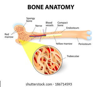
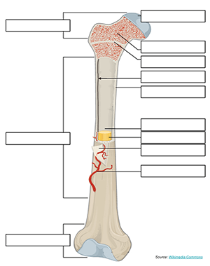
Label A Long Bone from www.biologycorner.com Above, you can see a labeled diagram showing the main bones of the body. Its upper end articulates with the tibia at the back of its head, whereas while attaching to the tibia with its lower end, it angles slightly forward. The epiphyseal plate is the area of growth in a long bone. Compact bone is organized as parallel columns, known as haversian systems, which run lengthwise down the axis of long bones. Below, you can find an unlabeled diagram ready for you to fill in yourself. Long, short, flat, irregular and sesamoid. They are one of five types of bones: Also, islands of red marrow may be seen anywhere in the skeleton, typically in a subcortical distribution, often with central yellow marrow giving it a bull's eye appearance on axial imaging.
Above, you can see a labeled diagram showing the main bones of the body.
In osseous tissue, bone cells are surrounded by a solid matrix of minerals and proteins. They also increase in width through appositional growth. Compact bone is organized as parallel columns, known as haversian systems, which run lengthwise down the axis of long bones. Below, you can find an unlabeled diagram ready for you to fill in yourself. It consists of "osseous tissue" made of "osteocytes," or bone cells. The mineralizing surface is calculated as the sum of doubly labeled plus half of singly labeled surfaces per bone surface (ms/bs). These columns are composed of lamellae, concentric rings of bone, surrounding a central channel, or haversian canal, that contains the nerves, blood vessels, and lymphatic system of the bone. May 31, 2021 · now that you know a little bit more about the types and locations of bones, why not test your memory with a bone labeling exercise? Also, islands of red marrow may be seen anywhere in the skeleton, typically in a subcortical distribution, often with central yellow marrow giving it a bull's eye appearance on axial imaging. The epiphyseal plate is the area of growth in a long bone. The long bones are those that are longer than they are wide. Above, you can see a labeled diagram showing the main bones of the body. Mineral apposition rate (mar) represents the mean speed at which individual osteoid seams are mineralized.